Ophthalmic Simulation Tools – Website Content
Advanced Clinical Calculators & Simulation Tools
Welcome to my comprehensive suite of ophthalmic simulation tools, designed to enhance clinical decision-making and patient care in modern eye care practice. These evidence-based calculators provide precise predictions and visualizations essential for refractive surgery planning, optical analysis, and astigmatism management.
These interactive tools leverage advanced algorithms to deliver accurate, real-time calculations that support your clinical expertise. Whether you’re planning a corneal procedure, analyzing wavefront aberrations, expanding depth of focus or managing complex astigmatism cases, these simulators offer the computational precision you need for optimal patient outcomes.
Corneal Surgery Residual Thickness Predictor
Ensuring Safe Surgical Outcomes
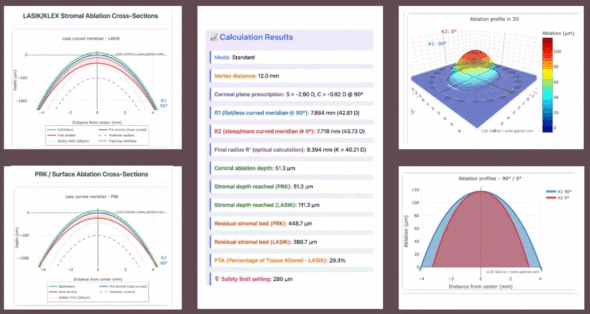
This advanced calculator predicts post-operative residual stromal thickness following corneal refractive procedures. By integrating pre-operative pachymetry data with planned ablation profiles, this tool helps surgeons maintain safe residual bed thickness thresholds, minimizing the risk of post-surgical ectasia. Essential for LASIK, PRK, and SMILE procedures, it provides instant visualization of tissue removal patterns and safety margins, supporting evidence-based surgical planning and enhanced patient safety protocols.
Key Features:
- Real-time thickness calculations
- Multiple surgical technique profiles
- Safety threshold alerts
- Customizable nomograms
AIOLsci – Patient-Specific IOL Performance Predictor
Personalized IOL Selection Through Advanced Optical Modeling
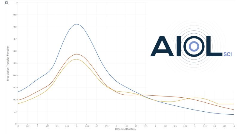
AIOLsci revolutionizes IOL selection by providing objective, patient-specific predictions of visual outcomes. This sophisticated platform bridges laboratory physics and clinical decision-making by combining empirically measured IOL wavefronts with personalized aspheric corneal models (R, Q). Unlike standard approaches that rely on average eye models, AIOLsci computes through-focus MTF to quantify exactly how a specific IOL will perform in your patient’s unique optical system. The platform features a comprehensive library of IOL designs based on proprietary optical bench measurements, capturing real manufacturing performance including asphericity, refractive profiles, and diffractive elements. Manufacturer-neutral and compatible with any biometer or topographer, AIOLsci delivers results through intuitive contrast curves and depth-of-field estimates, enabling direct comparisons between monofocal, EDOF, and multifocal options. This ensures each IOL choice is optimally tailored to the individual patient’s corneal optics rather than statistical averages, ultimately improving surgical outcomes and patient satisfaction in modern cataract surgery.
Key Features:
- Patient-specific corneal modeling (R, Q)
- Through-focus MTF analysis at multiple frequencies
- Empirically measured IOL database
- Manufacturer-independent comparisons
- Depth of field predictions
- Device-agnostic compatibility
Toric Phakic IOL Optimal Rotation Calculator
Minimize Residual Astigmatism in Phakic Lens Implantation
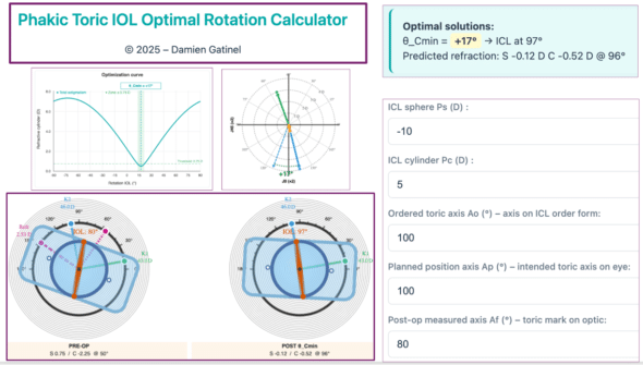
This interactive tool determines and visualizes the optimal rotation angle of a toric phakic intraocular lens (e.g., ICL, IPCL, or similar) to minimize postoperative residual astigmatism. It performs full vector analysis of corneal and lenticular astigmatism (J0/J45), converts toric power between the phakic IOL plane and corneal plane using the Distance between the corneal and phakic IOL plane (ELP equivalent), and displays optimization curves alongside frontal and surgical views. Designed as a decision-support and teaching tool for refractive surgeons performing phakic IOL implantation.
Key Features:
- Optimization of θ for minimal cylinder and target SE
- Phakic IOL→Cornea toric conversion with ELP (Gatinel Q-ratio and vergence)
- Frontal and surgical views, profiles and J0/J45 vector plots
- Dedicated to ICL, IPCL and similar phakic toric lenses
Wavefront Aberration to Retinal Image Simulator
Visualizing Visual Quality
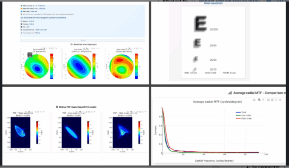
Transform complex wavefront data into meaningful clinical insights with our retinal image quality predictor. This sophisticated simulator converts Zernike polynomial coefficients and higher-order aberrations into visual representations of predicted retinal image quality. By modeling how optical aberrations affect point spread function and modulation transfer function, clinicians can better communicate expected visual outcomes to patients and optimize treatment strategies for aberration-correcting procedures.
Key Features:
- Zernike polynomial analysis up to 6th order
- PSF and MTF visualization
- Comparative pre/post treatment modeling
- Patient-specific pupil size adaptation
Toric IOL Optimal Rotation Calculator
Minimize Residual Astigmatism with Precise Rotation
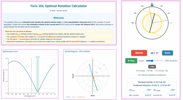
This interactive tool determines and visualizes the optimal rotation angle of a toric intraocular lens (IOL) to minimize postoperative residual astigmatism. It performs full vector analysis (J0/J45), converts toric power between the IOL plane and corneal plane using the Effective Lens Position (ELP), and displays optimization curves alongside frontal and surgical views. Designed as a decision-support and teaching tool for cataract surgeons.
Key Features:
- Optimization of θ for minimal cylinder and target SE
- IOL→Cornea toric conversion with ELP (Gatinel Q-ratio and vergence)
- Frontal and surgical views, profiles and J0/J45 vector plots
PEARL-DGS IOL Power Calculator
Open-Source AI-Based Formula for Complex Cases
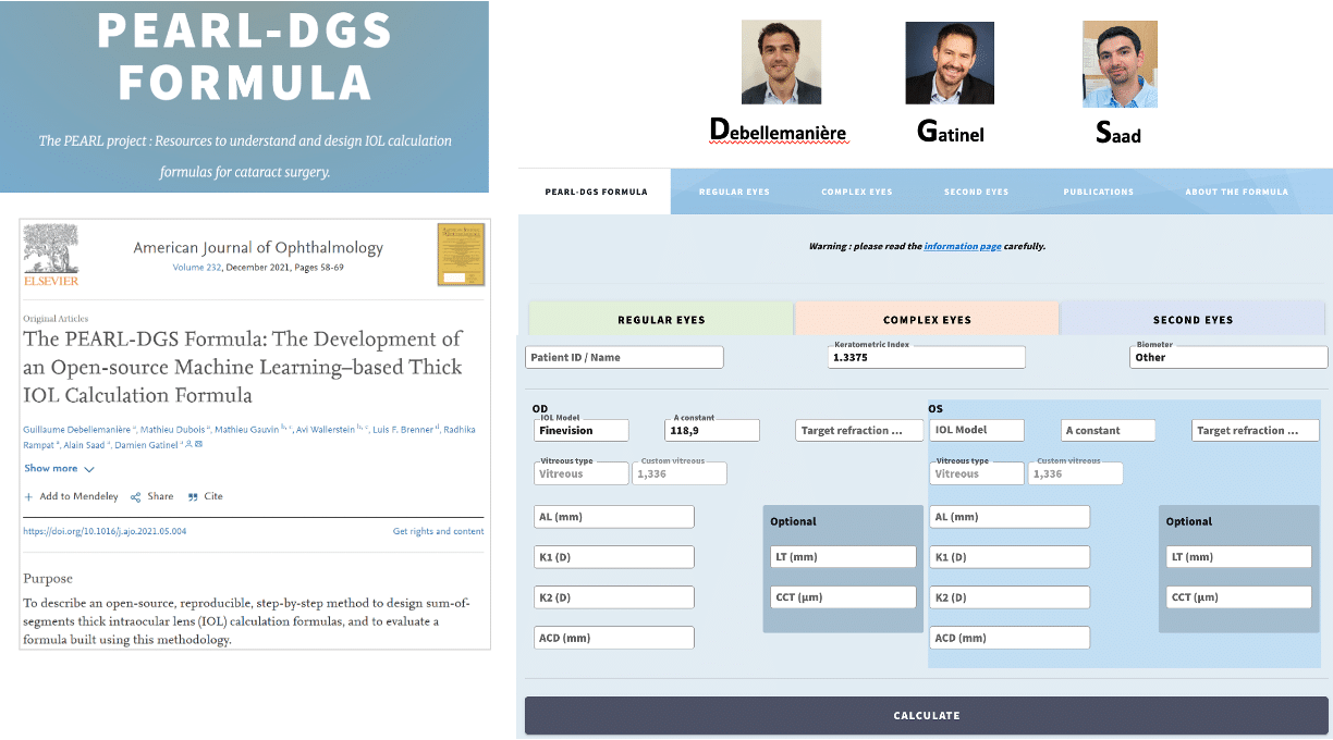
The PEARL-DGS (Postoperative spherical Equivalent prediction using ARtificial Intelligence and Linear algorithms) formula represents a breakthrough in IOL power calculation, combining thick lens equations with machine learning models to predict posterior corneal radius and the theoretical internal lens position (TILP). Developed by Debellemanière, Gatinel, Saad and published as open-source software, this advanced calculator features three specialized platforms. The « Regular Eyes » section handles standard cataract cases with customizable options for keratometric index, biometer type, and IOL model specifications, including support for meniscus designs. The « Complex Eyes » platform addresses challenging scenarios including post-refractive surgery eyes (myopic/hyperopic LASIK/PRK), radial keratotomy, scarred corneas, and ICL-implanted eyes, with automatic adjustments to lens position predictors and corneal power calculations. The « Second Eyes » section leverages first-eye surgical data to enhance prediction accuracy through back-calculation algorithms. Optional parameters including LT, CCT, and WTW measurements further refine predictions when available, making PEARL-DGS a comprehensive solution for achieving optimal refractive outcomes across the full spectrum of clinical presentations.
Key Features:
- Machine learning-based TILP prediction
- Post-refractive surgery algorithms
- Radial keratotomy and scarred cornea options
- ICL adjustment capabilities
- Second-eye optimization algorithms
- Open-source methodology
Combined Astigmatism Calculator & Visualizer
Mastering Complex Cylindrical Corrections
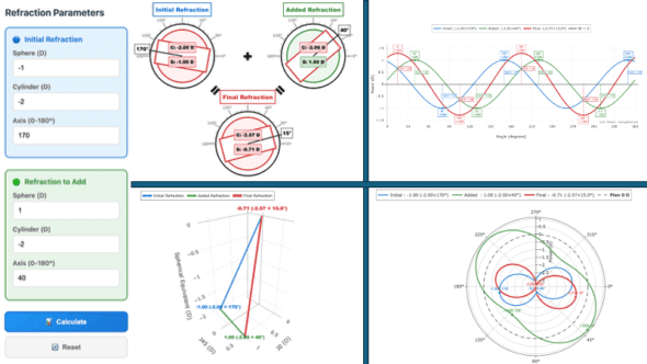
Navigate the complexities of astigmatism management with our comprehensive vector analysis tool. This calculator combines multiple astigmatic components—corneal, lenticular, and residual—providing both numerical and graphical representations of resultant cylinder power and axis. Ideal for toric IOL planning, crossed-cylinder calculations, and understanding obliquely crossed cylinders, this tool simplifies complex optical calculations while maintaining mathematical precision.
Key Features:
- Vector addition and subtraction
- Double-angle plot visualization
- 2D and 3D vectorial visualization
- Sinusoidal modeling visualization
Wavefront to Radial Vergence Power Map Converter
From Microns to Diopters – Clinical Translation
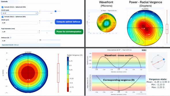
Bridge the gap between wavefront analysis and clinical refraction with this powerful conversion tool. Transform Zernike polynomial coefficients expressed in microns into clinically interpretable refractive power distribution maps in diopters. Unlike traditional wavefront displays showing optical path differences, this simulator generates radial vergence maps that directly reveal myopic and hyperopic zones across the pupil aperture. This intuitive visualization provides immediate clinical relevance, allowing practitioners to correlate aberrometry findings with manifest refraction measurements and better understand the refractive impact of higher-order aberrations on visual performance.
Key Features:
- Zernike-to-diopter conversion algorithms
- Radial vergence power mapping
- Myopic/hyperopic zone identification
- Direct correlation with clinical refraction
Corneal Asphericity Impact Analyzer
Exploring Q-factor Effects on Optical Performance
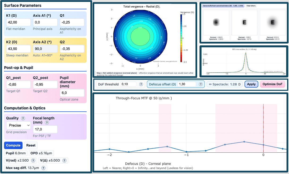
Dive deep into the complex relationship between corneal asphericity (Q-factor) and optical performance with this comprehensive simulation tool. This advanced analyzer demonstrates how variations in corneal asphericity directly influence spherical aberration (Z4,0), vergence distribution patterns, and the induction of useful depth of focus. Through interactive 3D visualizations and real-time calculations, clinicians can explore how changing Q-values affect wavefront aberrations, visualize the resulting intrapupillary power distribution (induced multifocality), and quantify the impact on through-focus visual performance. Essential for presbyopia-correcting procedures and aspheric IOL selection, this tool bridges theoretical optics with clinical applications by illustrating the delicate balance between corneal shape and visual quality.
Key Features:
- Interactive Q-factor manipulation with real-time aberration analysis
- Radial and Laplacian vergence mapping for multifocality assessment
- Through-focus MTF analysis with depth of focus optimization
- PSF visualization and convolved letter simulation
- Spectacle and corneal plane conversion capabilities
- Integration of native ocular spherical aberration modeling
Elevate Your Clinical Practice
Experience the precision and efficiency of professional-grade ophthalmic calculators. Each tool is continuously updated to reflect the latest clinical research and surgical techniques.
Click on any image to access each simulator directly.
Les commentaires sont fermés.
