This paper explores the topography of the corneal epithelium and Bowman layer, which is the most superficial layer of the corneal stroma, in normal myopic eyes.
Link toward the JCRS paper (valid until October 2016)
The Bowman layer is a constitutive layer of the cornea that separates the corneal epithelium from the anterior stroma. Routine corneal topographic or OCT examinations are performed using topographic analysis of the air–tear film interface, which delineates the anterior surface of the corneal epithelial layer. The thickness of the epithelial layer is not constant over the entrance pupil, and its refractive index differs from the underlying corneal tissue.
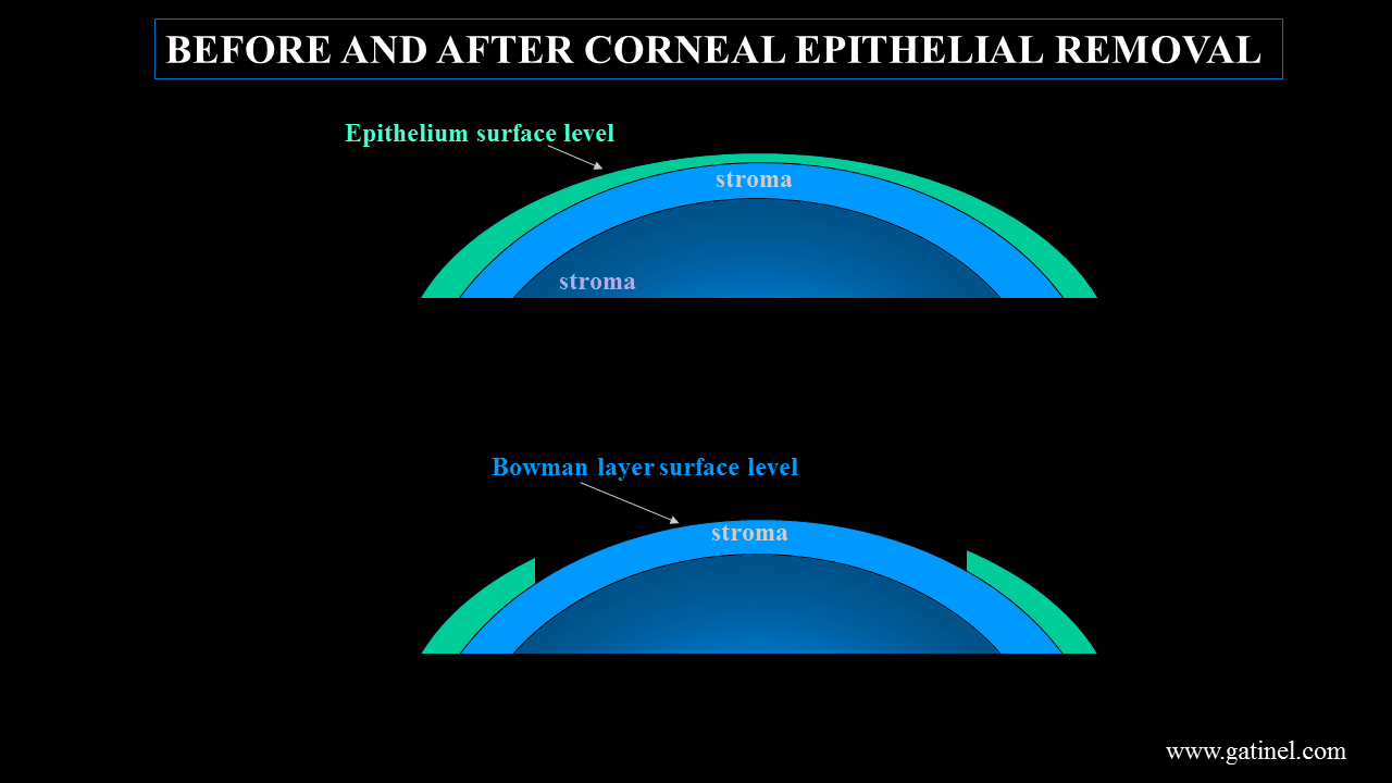
Hence, the epithelial layer may induce subtle changes to the topography of the underlying corneal stroma. Simply removing the epithelium may alter the refractive and geometric properties of the cornea prior to photoablation.
Conventional or customized photoablative techniques used to modify the optical power of the cornea achieve their effect by altering the anterior corneal surface contour. In customized surface ablation techniques, the preoperative wavefront and/or corneal topography measurements are performed with the epithelial layer in place, while the laser ablation is performed on the underlying stroma (Bowman layer) after epithelial removal. How important is the change caused by the removal of corneal epithelium?
The topography of the Bowman membrane can bring relevant information regarding the properties of the corneal epithelium layer, by comparing the topographies obtained before and after epithelial removal.
In 2007, we were the first to analyze the effect of epithelial removal during a PRK procedure in refractive surgery candidates. Our results confirmed that in photorefractive keratectomy (PRK), the simple removal of the corneal epithelium can modify the geometric (and therefore the refractive) properties of the cornea before excimer ablation. Apart from this study which was performed with a slit scanning topograph (Orbscan IIz), the topography of Bowman layer has not been extensively studied in vivo, in normal eyes of myopic patients.
The aim of this study was to further explore the Bowman’s layer shape by comparing the front surface specular corneal topography and the Bowman’s membrane specular topography in normal eyes. In this survey, we used a Placido topographer device (OPD-Scan II ®, Nidek, JAPAN), to measure several outcomes of the anterior cornea before and after epithelial removal during a standard Photo Refractive Keratectomy (PRK).
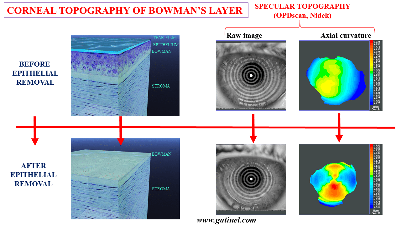
The preoperative OPD-Scan topography was performed on each eye 10 minutes before the PRK and administration of topic anesthesia. Using a 20% alcohol solution applied over the corneal surface during 20 seconds, the epithelium was removed on the 8 central corneal millimetres. The corneal surface was then rinsed with a balanced salt solution and dried with cellulose sponges. Immediately after, three postoperative topographies were performed. The examiner checked each image quality before recording it. Patient returned to the surgical bed and excimer laser procedure were performed as conventional.
The obtained specular image of the denuded allowed to compute the topography of the Bowman membrane in normal myopic eyes.
We present the results obtained on 90 myopic eyes of 51 patients (36 men, mean age 32.2 years, ranging from 20.5 to 56.3 years).
The topography of Bowman layer was significantly steeper than that at the surface of the epithelium in low to moderate myopic eyes. The epithelial layer tended to decrease the magnitude astigmatism and prolateness of Bowman layer.
The following figure represents the vector change (magnitude and axis) after epithelial removal. On average, the Bowman layer exhibits increased with the rule toricity (astigmatism), in comparison to the epithelial layer.
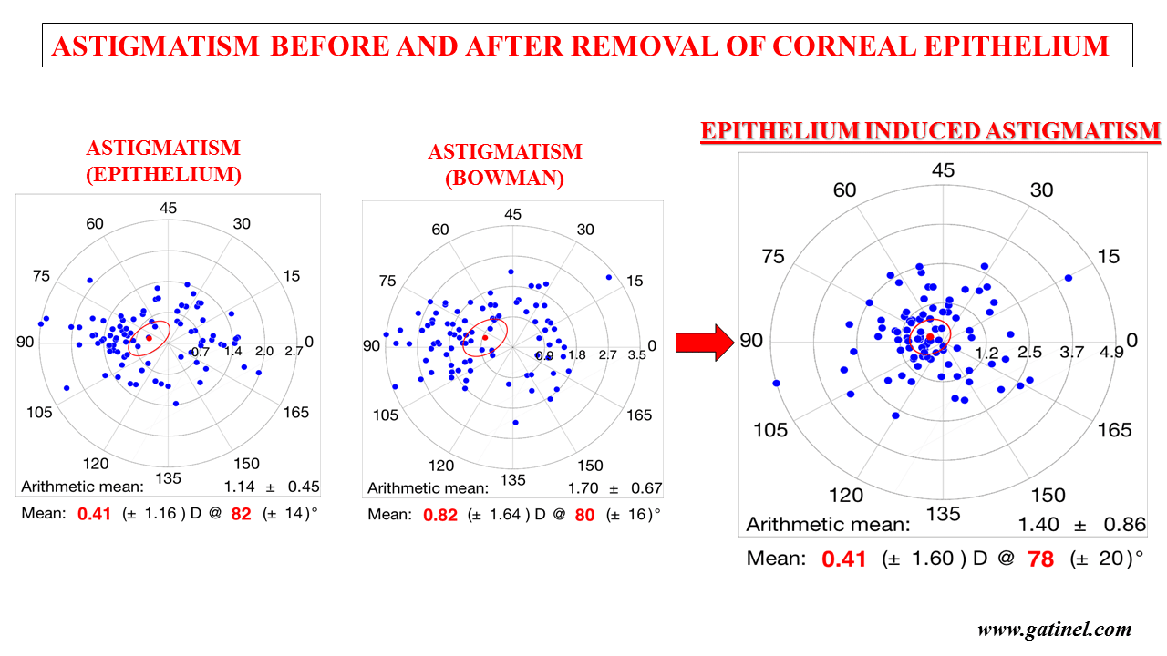
The magnitude of these changes are optically significant for the level of precision required in refractive surgery.