Eye rubbing: a sine qua non for keratoconus? D Gatinel. Int K Kerat Ect Cor Dis, 2016;5(1):6-12
(Link to the original article here) download : Eye rubbing a sine qua non for keratoconus

Eye rubbing as the root cause of keratoconus?
Keratoconus has been labelled a “dystrophy of unknown origin”, and I have long been fascinated by this mysterious disease. A large proportion of my clinical practice is dedicated to the diagnosis, management and prevention of keratoconus, and for many years I have been intrigued by how structural changes and deformation of the corneal wall are so pronounced in keratoconus, yet paradoxically, few detectable genetic and molecular abnormalities exist in this condition.
Eye rubbing has long been acknowledged as a risk factor for keratoconus, but I believe its role in the pathogenesis of keratoconus has not been accorded sufficient prominence. I have thus undertaken the task to pen an article to suggest to readers a pathophysiological concept of keratoconus that I am increasingly convinced of: that eye rubbing is the root cause or sine qua non for keratoconus (article download). As such, eye rubbing is not a « risk factor », as often coined in medical litterature, but the direct cause of a syndrome marked by a corneal deformation labeled « keratoconus ».
In this article, I have put forth the theory that keratoconus is not a dystrophy of unknown genetics and biomolecular substratum, but rather a syndrome caused by eye rubbing. i.e what has been called “keratoconus” is in essence the direct consequence of mechanical trauma to the cornea by chronic and incessant eye rubbing, resulting in the progressive deformation and thinning of the corneal wall which are hallmarks of the disease. Eye rubbing is the main character of the « mechanical » hypothesis as the etiology of keratoconus:
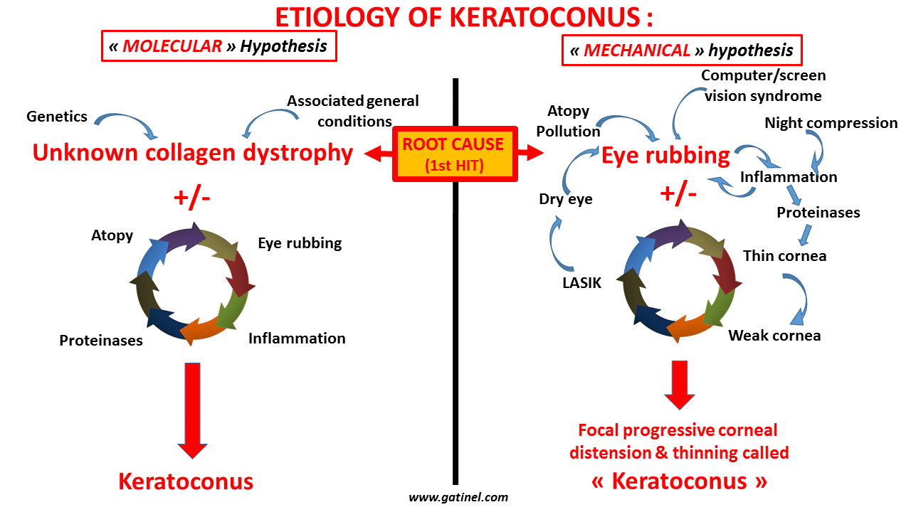
The Molecular Hypothesis vs the Mechanical Hypothesis illustrated in a schematic diagram. The Mechanical Hypothesis states that keratoconus is primarily caused by eye rubbing. Eye rubbing is mainly triggered by atopy, air pollution, and extended computer screen viewing. In contrast to the Molecular Hypothesis where genetics, environmental and other unknown general factors are keys to the apparition of the disease, the Mechanical Hypothesis simply states that the structural corneal changes and deformation are initiated and aggravated by a mechanical cause: eye rubbing. Additional mechanical factors, such as corneal refractive surgery or night compression of the cornea (by pressure of the hand or pillow) may also accelerate the corneal deformation. The prolonged contact between the eye and eyelids with the pillow of mattress can cause local contamination and irritation. This, in turn, leads to local itchness, and triggers eye rubbing, expecially in the morning. This explains the striking correspondence between the » head side » on which patients sleep and the topographic features evocative of keratoconus.
Eye rubbing can cause inflammation. The effect of the mechanical stress (distension) is further accentuated by the release of proteinases in the stroma, explaining the progressive thinning of the cornea, which in turn makes it more vulnerable to the trauma caused by rubbing. LASIK cause eye-dryness, and this can in turn trigger eye rubbing, on a more vulnetable (thinned by the refractive surgery procedure).
In the Mechanical Hypothesis, keratoconus cannot occur without a repeated mechanical injury such as eye rubbing. When the duration and frequency of eye rubbing exceed the native structural and biomechanical resistance capacity of the cornea, the mechanical imbalance causes the cornea to deform, leading to characteristic topographical patterns, encompassing minor forms of deformation (keratoconus « forme fruste ») to the end stage KC. In my experience, the latter is always encountered in patients who rub the affected eye vigorously and frequently.
Keratoconus as a primary mechanical disease
Just as excessive ligament extension can cause a sprained ankle, chronic eye rubbing can cause the corneal collagen fibers to lose part of their biomechanical resistance, resulting in macroscopically obvious structural deformation. This biomechanical mechanism could also better explain the frequent disparity in the degree of affliction between the right and left eyes (patients frequently rub one eye more often and more vigorously than the other) and the focal nature of keratoconus, which has been recently evidenced.
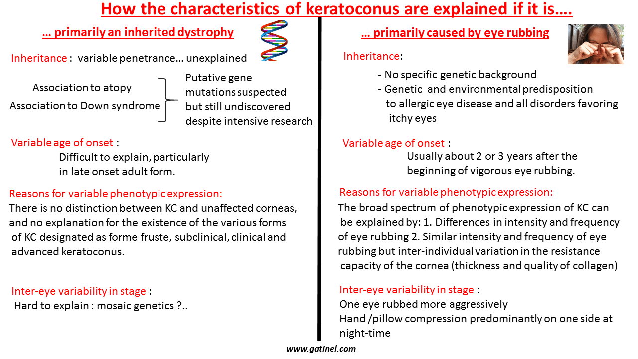
The exact genetics of keratoconus has yet to be elucidated. The frequency of occurrence in close family members is not clearly defined and is estimated to be less than 20%. In the ‘eye rubbing as a sine qua non for keratoconus’ hypothesis, the influence of genetics is related to the predisposition to conditions that lead to increased eye rubbing, and to corneal thickness and resistance. Down syndrome and atopy are obviously such conditions. Sleep apnea has also been associated with an increased incidence of keratoconus. The deprivation of good quality sleep causes chronic fatigue, and fatigue can induce patients to rub their eyes more frequently. I have often observed cases of late onset keratoconus (after 30 years of age) in workers who had a disturbance of their biological clock by a shift in their work hours from day to night, causing them to be chronically fatigued, and inducing them to rub their tired eyes frequently. A recent male predominance has been discovered in KC. In my experience, women who wear eye make-up tend to rub their eyes less often and less aggressively than men. However, some women with keratoconus tend to rub their eyes heavily after make-up removal. Dry eye and ocular irritation are frequent during pregnancy: the occurrence of keratectasia after pregnancy may be explained again by increased eye rubbing. It is easier to explain the variation in the age of presentation, laterality, severity and broad spectrum of phenotypic expression by excessive eye rubbing than a corneal degenerescence caused by an unknown genetic disorder or molecular cascade. For the same eye rubbing intensity, duration and frequency, corneas with natively reduced thickness and biomechanical resistance may deform more readily and significantly than thicker and stronger corneas. Exposing populations with thinner corneas to high level of pollution, dry air, irritating or allergic agents and bad and/or intensive working conditions may account for the prevalence of keratoconus in some socio-ethnic groups. The recent increase in computer usage has been linked to various ocular symptoms gathered together to be called « Computer Vision Syndrome », and this includes eye fatigue, which elicits eye rubbing, which may in turn account for the increase in keratoconus prevalence.
Marfan syndrome vs Keratoconus
These and other concepts are explored further in the article, through both scientific and anecdotal evidence. A pertinent comparison with the cornea changes in Marfan Syndrome is also made:
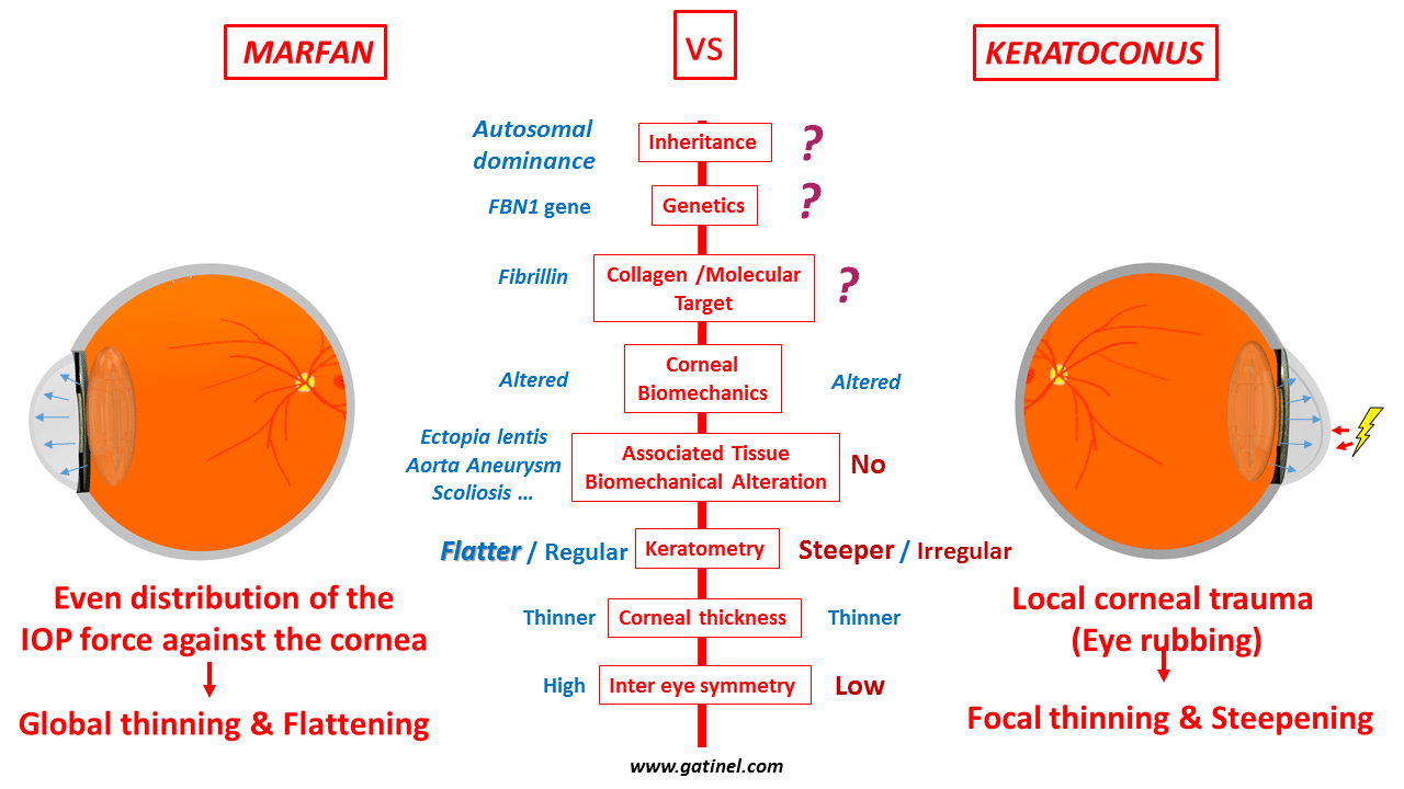
Marfan syndrome is a genetic disorder of connective tissue. It is caused by a mutation in the gene controlling a protein called fibrillin-1, which is widely distributed in connective tissue throughout the body, and is a major component of collagen fibrils. In this syndrome, many organs of the body are affected, including the eye. The aorta, which media comprises large layers of connective tissue, can progressively weaken and stretch under pressure from within the blood vessel, leading to a bulge in the vessel wall called an « aneurysm ». In the eye, Marfan syndrome is associated with a displacement of the crystalline lens (ectopia lentis), due to weakening and loss of resistance of the zonular fibers which attach the lens to the sclera. These zonular fibers are partly made up of fibrillin-1. The lens subluxation caused by the broken or attenuated zonular fibers is typically bilateral, symmetric, and in the superotemporal direction, although it may occur in other directions.
Marfan syndrome represents a perfect counter-example to explain the irrelevance of current theories on the pathogenesis of keratoconus, which describe keratoconus to be an unknown collagen dystrophy associated with environmental, cellular and genetic factors causing a degenerative change in the cornea. In fact, based on these assumptions, Marfan syndrome should perfectly support the theories of keratoconus that attempt to explain the ectatic process. In Marfan syndrome, the gene mutation is identified, and the fibrillin-1 molecule involved in this connective tissue dystrophy is responsible for the reduction of the strength of the collagen present in the ocular tissues, including the corneal stroma. Yet, despite all these features, no keratoconic or ectatatic pattern is seen in the corneas of patients with Marfan syndrome. The Marfan corneas are thinner, but tend to be flatter instead of steeper. This surprising topographical feature can be easily accepted a priori if one considers that in normal conditions, the main force exerted against the cornea is intraocular pressure. Apparatus for measuring pressure within an eye typically include mechanisms to increasingly deform the cornea by applying a progressively increasing force onto it. This force (eg: a puff of air) is responsible of an inward deformation of the corneal surface, which, for the same intraocular pressure, will be easier if the cornea is thinner or biomechanically weaker. Because the force resulting from intraocular pressure is evenly distributed against the posterior surface of the cornea and the inner surface of the sclera, a softer eyeball will undergo a progressive distension of its shell, which causes the local radii of curvature of the cornea to increase (and the corneal curvature to decrease), with concomitant progressive thinning. Hence, in the absence of a localized or focal additional force or trauma, the biomechanical weakening of Marfan corneas results in a flatter corneal surface. This distension of the eyeball also contributes to its increase in axial length, and most Marfan patients indeed suffer from axial myopia . Abnormally flat corneas and axial myopia both correspond to the ocular diagnostic criteria for Marfan syndrome.
Although a flat keratometry is a diagnostic criteria for a Marfan cornea, some association between Marfan syndrome and keratoconus has been reported. This however does not contradict the mechanical theory: the biomechanically weaker corneas of Marfan patients may be more vulnerable to the effects of eye rubbing.
In contrast to the Marfan cornea, the typical topographic pattern of a keratoconus cornea includes steepening and asymmetry of the corneal surface and changes in the corneal thickness, including central or paracentral accelerated thinning. Invoking an external source of trauma to the cornea (such as eye rubbing) as a pathogenetic cause provides a better explanation to these findings than still unraveled processes of molecule change mysteriously affecting only the cornea and preserving the other tissue components of the body. The force exerted against the corneal shell by the pressure of the fingers, particularly the knuckles, may be considerable. Repetitive force can cause stretching and disorganization of the corneal collagen lamellae. An acute rise in intraocular pressure from sudden reduction in eye volume from the compression of the globe by the fingers could also stretch the scleral shell and lead to an axial myopic refractive shift. Despite contradicting reports, the axial length of keratoconus patients seems to be slightly but significantly higher than in non-keratoconus patients.
There will understandably be skeptics and naysayers to this theory, especially since the hypothesis may be impossible to prove. However, the primary purpose of this article is to increase the awareness of the potential ill-effects of eye rubbing and its association with keratoconus. Highlighting its causative role may help to dramatically reduce the incidence of keratoconus, and stop its progression in eyes already affected.
It is interesting to note that conditions that are characterized by the presence of corneal inflammation generally induce a reduction in corneal curvature, not an increase in corneal curvature. This also underlines the importance of the mechanical hypothesis to account for the focal steepening which is observed in corneas with keratoconus.
In conclusion:
The “eye rubbing as a sine qua non for keratoconus” theory blatantly defies the widely accepted concept of keratoconus being a corneal dystrophy of unknown origin. To many, it may appear provocative in its simplicity, and on a personal level, would have triggered some skepticism 6 or 7 years ago. However, in recent years, after conceiving the hypothesis and applying it to my day to day management of keratoconus patients, the skepticism is waning. The theory is compatible with most of what is known about keratoconus. In fact, day by day, after each patient interview, I am increasingly convinced that this mechanical first hit theory is the answer to the enigma surrounding keratoconus. This opens fascinating perspectives. It could close the chapter of the mystery of the pathogenesis of keratoconus opened by Dr John Nottingham’s 1854 treatise on the conical cornea. In theory, it could also lead to the eradication the disease if it were possible to prohibit everyone from chronically and incessantly rubbing their eyes. However, from a practical standpoint, this is probably an impossible mission.
A less ambitious but important goal is still attainable, and this involves reducing the incidence and progression of keratoconus by increasing the awareness of patients on the potential dangers of chronic and vigorous eye rubbing, and encouraging them to refrain from it. In the management of my many patients with keratoconus, I religiously counsel them on the dangers of eye rubbing, and tell all of them to stop rubbing their eyes. I also routinely perform detailed evaluation of their condition, including refraction and subtractive topography maps between each consultation to document disease progression. None of my patients who have completely stopped rubbing their eyes have seen progression in their disease since they kicked the habit. These observations are still ongoing, and will require a longer follow-up, but if they stand the test of time, they will bring forth a strong argument to support the “eye rubbing as a sine qua non for keratoconus’ theory, and more importantly, demonstrate its clinical benefits.
The wear of the mask should be fine, the force exerted against the cornea is negligible compared to that of the rubbing knuckles!
Good day dr. Gatinel
I have been diagnosed with a mild posterior blepharitis one year after doing a femto-lasik intervention to correct myopia. Among other things, the doctor recommended I do warm compresses with a heated eye mask (Bruder), twice a day for 10 minutes.
My question is this: after having read all of your articles on the link between rubbing and post-Lasik ectasia, do you think that wearing the mask twice a day as prescribed (without doing a lid massage afterwards) puts enough pressure on the eye to increase the risk of Ectasia? How much of a pressure are we talking about that would represent a risk factor? Many thanks
Thank you. Although there are many papers that point eye rubbing as a « risk factor » (where as you can read here, I consider it as the direct cause of the deformation), I am not aware of a well conducted experimental study that would aim at investigating the effect of the eye rubbing forces on the corneal architecture. I presume that the shearing forces would weaken the collagen fibers adhesions and create a focal paracentral weakening. This eventually leads to the buckling that is in my humble opinion the true nature of keratoconus. I would be interested to cooperate in such study. A mathematical modeling could aslo work.
Excellent analysis,dr Gatinel, it will more interesting to reproduce the mechanical stress of rubbing in experimental conditions and study corneal architictural changes. I dont know if there is any study in this topic
This is a great paper, your hypothesis is fascinating and mind opening, … All this makes as lot of sense. Thanks.
[…] Eye rubbing as the root cause of Keratoconus? […]
Merci pour vos commentaires et effectivement, vous semblez avoir compris que les frottements oculaires ont provoqué et aggravé le kératocône, qui est logiquement stable depuis que vous avez cessé de frotter vos yeux. Comme stipulé dans cet article, et c’est un argument fort en faveur de l’effet initiateur des frottements, tous mes patients, suivis régulièrement, qui ont cessé de se frotter les yeux, sont stables et ne progressent plus. Je m’occupe du dépistage, du suivi du kératocône mais l’adaptation en lentilles rigides requiert des compétences spéciales en matière de contactologie. Aussi, il est préférable que vous preniez directement rdv avec un spécialiste adaptateur de lentilles pour le KC.
Bonjour Docteur,
Je pense que vous avez mis le doigt sur la cause principale du KC, moi-même ayant eu l’habitude de souvent frotter mes yeux dans mon enfance j’ai développé un KC bilatéral qui est stable depuis plusieurs années maintenant depuis que je ne me frotte quasiment plus les yeux (j’avais fait le lien également).
J’ai appelé votre secrétariat pour prendre rdv avec vous pour un suivi et une adaptation de lentille (le Pr Cochet qui me suivait n’exerce malheureusement plus après sa retraite bien méritée), mais l’on m’a dit que je ne pouvais avoir de rdv qu’avec un autre médecin et non vous en particulier, ce que je souhaitais car pour moi c’est vous le spécialiste du KC…
Je retenterai sûrement bientôt :)
Merci pour vos travaux très intéressants !
Bien cordialement